Coding and Documentation Guidance
Written by: AIHC Blogger
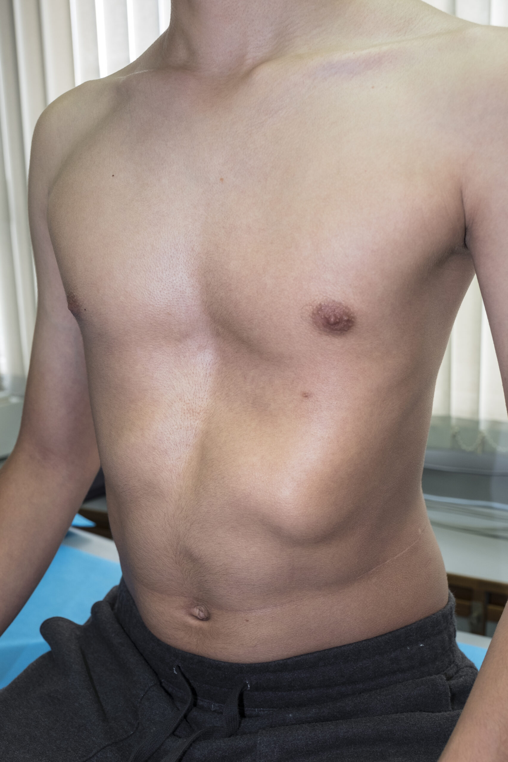
Pectus excavatum is a Latin term that means “hollowed chest.”
Pectus excavatum is a congenital condition due to too much growth of the cartilage that joins the ribs to the sternum causing the sternum to grow inward. As the cartilage pushes the sternum inward, the child’s chest appears to be sunken. The condition is also called “sunken chest” or “funnel chest.”
According to Boston Children’s Hospital, there is no known cause for pectus excavatum. Some studies investigating a genetic component are underway. Although the majority of cases don't involve a family history, there are many that do — enough to warrant suspicion that genes may play a significant role.
This condition can present in toddlers but typically becomes apparent during a growth spurt and puberty.
Pectus excavatum may be mild, moderate or severe. Even mild cases of pectus excavatum can make children feel self-conscious about their appearance. The way the chest looks may cause emotional stress for the child. If the condition is severe, the heart and lungs can be affected. The lower half of the breastbone may press on the heart and lungs. This can cause shortness of breath with exercise.
Many children with pectus excavatum don’t require any treatment at all because their condition is so mild. However, in a more severe case, the sternum can press on the heart and lungs, but the effects on the heart and lungs are usually minor and typically only occur with extreme exercise.
The symptoms of pectus excavatum depend on when the condition is diagnosed. In infancy, symptoms of pectus excavatum can include:
- Hollow depression in the chest that may be broad and shallow, deep and narrow, or irregular more rapid breathing than normal
The symptoms can be both physical and psychological. Physical symptoms can include:
- Shortness of breath with exercise;
- Recurrent respiratory infections;
- Decreased stamina compared to peers;
- Fatigue;
- Pain – in chest and back areas which tends to come and go (may be worse during periods of excessive growth);
- Poor posture;
- Lateral curvature of the spine, absence of the curve of the upper back, hooked shoulders and a broad thin chest;
- Palpitations, irregular heartbeat.
The psychological impact for teens and adults can be significant, especially regarding self-esteem and confidence. Symptoms can include:
- Significant embarrassment from the appearance of the chest;
- Self-esteem issues;
- Withdrawn behavior;
- Avoidance of certain activities (where chest may be exposed);
- Clinical depression.
How It Is Diagnosed & Treated
This condition is usually diagnosed during a physical examination and can be diagnosed via a visual examination of the anterior chest wall. Evaluation needs to be individually tailored to the patient's symptoms. It typically affects 4-5 ribs on each side of the sternum. The condition’s visibility varies from one case to the next, and there can sometimes be significant asymmetry between the two sides of the chest.
Pectus excavatum is also a common symptom of Marfan syndrome, and many children with spinal muscular atrophy (SMA) develop PE due to the diaphragmatic breathing that commonly accompanies that disease.
Diagnostic tests typically ordered are X-rays, MRI or CT scan of the chest. Other tests commonly ordered are:
- Electrocardiogram (EKG or ECG)
- Echocardiogram (cardiac ultrasound)
- Pulmonary function test (PFT)
- Exercise function test or cardiopulmonary exercise testing (CPET)
In severe cases, repair of a pectus excavatum may help the function of the heart and lungs. But in most cases, the benefits are mostly psychological.
The Nuss or Ravitch procedures are surgical treatment options. The goal of pectus excavatum repair is to relieve pressure on the heart and lungs that may impair function. This typically leads to improvements in breathing, exercise intolerance and chest pain. It is not uncommon for patients with pectus excavatum to feel as if their breathing and stamina are normal before surgery and then realize they feel much improved following correction.
Nuss procedure (repair w/metal pectus bar)
This minimally invasive procedure uses small incisions placed on each side of the chest. Long-handled tools and a narrow fiber-optic camera are inserted through the incisions. A curved metal bar is threaded under the depressed breastbone, to raise it into a more normal position. In some cases, more than one bar is used. The bars are removed after two or three years.
Ravitch technique (open repair)
This older procedure involves a much larger incision down the center of the chest. The surgeon removes the deformed cartilage attaching the ribs to the lower breastbone and then fixes the breastbone into a more normal position with surgical hardware, such as a metal strut or mesh supports. These supports are removed after 12 months.
21740 Reconstructive repair of pectus excavatum or carinatum; open
21742 Reconstructive repair of pectus excavatum or carinatum; minimally invasive approach (Nuss procedure), without thoracoscopy
21743 Reconstructive repair of pectus excavatum or carinatum; minimally invasive approach (Nuss procedure), with thoracoscopy
When removal of hardware is required, code 20680 is reported:
20680 Removal of implant; deep (e.g., buried wire, pin, screw, metal band, nail, rod or plate)
Diagnosis Code for Pectus Excavatum
The code for pectus excavatum falls under the ICD-10-CM category code Q67 for congenital musculoskeletal deformities of head, face, spine and chest.
Q67.6 Pectus excavatum
Congenital funnel chest
Additional Learning for Coders & Auditors
The National Library of Medicine (NIH) has updated their Pectus Excavatum continuing education activity as recently as July 2022. Click Here to learn more and access additional references on this topic.
Register for the Auditing for Compliance online training program w/option to certify.